Figure 1
Membranous nephropathy complicating relapsing polychondritis: A case report
Christopher Rice, Vatsalya Kosuru, John Jason White, Christine Van Beek, Rachel Elam*, Michael Clemenshaw, Laura Carbone and Leighton James
Published: 07 October, 2021 | Volume 5 - Issue 3 | Pages: 084-087
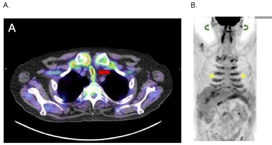
Figure 1:
F-18 Fluorodeoxyglucose (FDG) Positron Emission Tomography-Computed Tomography Imaging Demonstrating Diffuse Cartilaginous Inflammatory FDG Uptake. A. Axial fused Positron Emission Tomography-Computed Tomography (PET-CT): Anterior tracheal wall thickening with severe luminal narrowing and diffuse increased FDG uptake (red arrow). B. Anterior Whole Body Maximum Intensity Projection FDG PET-CT Image: Diffuse increased FDG uptake in the costal cartilage (yellow stars) and auricles (green curved arrows).
Read Full Article HTML DOI: 10.29328/journal.jcn.1001080 Cite this Article Read Full Article PDF
More Images
Similar Articles
-
Acute Tubulointerstitial Nephritis due to Phenytoin: Case ReportNilzete Liberato Bresolin*,Pedro Docusse Junior,Maria Beatriz Cacese Shiozawa,Marina Ratier de Brito Moreira,Natalia Galbiatti Silveira. Acute Tubulointerstitial Nephritis due to Phenytoin: Case Report. . 2017 doi: 10.29328/journal.jcn.1001004; 1: 019-025
-
Equine Anti-Thymocyte Globulin (ATGAM) administration in patient with previous rabbit Anti-Thymocyte Globulin (Thymoglobulin) induced serum sickness: A case reportJoseph B Pryor*,Ali J Olyaei,Joseph B Lockridge,Douglas J Norman. Equine Anti-Thymocyte Globulin (ATGAM) administration in patient with previous rabbit Anti-Thymocyte Globulin (Thymoglobulin) induced serum sickness: A case report. . 2018 doi: 10.29328/journal.jcn.1001013; 2: 015-019
-
A case report of Hypocomplementemic urticarial vasculitic syndrome presenting with Renal failureAmy M Hopkins,Angela M Gibbs,Ryan S Griffiths,Rupali S Avasare,Firas G Khoury*. A case report of Hypocomplementemic urticarial vasculitic syndrome presenting with Renal failure. . 2018 doi: 10.29328/journal.jcn.1001017; 2: 039-043
-
Lessons from the success and failures of peritoneal Dialysis-Related Brucella Peritonitis in the last 16 years: Case report and Literature reviewMuhammad Bukhari*,Azharuddin Mohammed,Abdullah Al-Guraibi,Ahmed Shahata,Mansour Al-Ghamdi. Lessons from the success and failures of peritoneal Dialysis-Related Brucella Peritonitis in the last 16 years: Case report and Literature review. . 2018 doi: 10.29328/journal.jcn.1001020; 2: 057-061
-
Anti-glomerular basement membrane disease: A case report of an uncommon presentationNatália Silva*,Luís Oliveira,Mónica Frutuoso,Teresa Morgado. Anti-glomerular basement membrane disease: A case report of an uncommon presentation. . 2019 doi: 10.29328/journal.jcn.1001027; 3: 061-065
-
Granulomatosis with polyangiitis (GPA) in a 76-year old woman presenting with pulmonary nodule and accelerating acute kidney injuryNoor Sameh Darwich*,Melissa Schnell,L. Nicholas Cossey. Granulomatosis with polyangiitis (GPA) in a 76-year old woman presenting with pulmonary nodule and accelerating acute kidney injury. . 2020 doi: 10.29328/journal.jcn.1001048; 4: 001-006
-
Unusual presentation of oxalate nephropathy causing acute kidney injury: A case reportAnas Diab*,Michelle M Neuman,Kareem Diab,Daniel Gordon. Unusual presentation of oxalate nephropathy causing acute kidney injury: A case report. . 2020 doi: 10.29328/journal.jcn.1001063; 4: 077-079
-
Prostate cancer-associated thrombotic microangiopathy: A case report and review of the literatureHHS Kharagjitsing*,PAW te Boekhorst,Nazik Durdu-Rayman. Prostate cancer-associated thrombotic microangiopathy: A case report and review of the literature. . 2021 doi: 10.29328/journal.jcn.1001068; 5: 017-022
-
Membranous nephropathy complicating relapsing polychondritis: A case reportChristopher Rice,Vatsalya Kosuru,John Jason White,Christine Van Beek,Rachel Elam*,Michael Clemenshaw,Laura Carbone,Leighton James. Membranous nephropathy complicating relapsing polychondritis: A case report. . 2021 doi: 10.29328/journal.jcn.1001080; 5: 084-087
-
Persistent symptomatic hyponatremia post-COVID 19: case reportHaifa Alshwikh,Ferial Alshwikh,Halla Elshwekh*. Persistent symptomatic hyponatremia post-COVID 19: case report. . 2022 doi: 10.29328/journal.jcn.1001090; 6: 058-062
Recently Viewed
-
Update on Mesenchymal Stem CellsKhalid Ahmed Al-Anazi*. Update on Mesenchymal Stem Cells. J Stem Cell Ther Transplant. 2024: doi: 10.29328/journal.jsctt.1001035; 8: 001-003
-
Impact of Chronic Kidney Disease on Major Adverse Cardiac Events in Patients with Acute Myocardial Infarction: A Retrospective Cohort StudyAbbas Andishmand, Ehsan Zolfeqari*, Mahdiah Sadat Namayandah, Hossein Montazer Ghaem. Impact of Chronic Kidney Disease on Major Adverse Cardiac Events in Patients with Acute Myocardial Infarction: A Retrospective Cohort Study. J Cardiol Cardiovasc Med. 2024: doi: 10.29328/journal.jccm.1001175; 9: 029-034
-
Myxedema Coma and Acute Respiratory Failure in a Young Child: A Case ReportRinah Elaisse R Dolores, Marion O Sanchez*. Myxedema Coma and Acute Respiratory Failure in a Young Child: A Case Report. Ann Clin Endocrinol Metabol. 2023: doi: 10.29328/journal.acem.1001027; 7: 008-013
-
A General Evaluation of the Cellular Role in Drug Release: A Clinical Review StudyMohammad Hossein Karami* and Majid Abdouss*. A General Evaluation of the Cellular Role in Drug Release: A Clinical Review Study. Clin J Obstet Gynecol. 2024: doi: 10.29328/journal.cjog.1001162; 7: 042-050
-
Are Biofungicides a Means of Plant Protection for the Future?Radek Vavera*, Josef Hýsek. Are Biofungicides a Means of Plant Protection for the Future?. J Plant Sci Phytopathol. 2024: doi: 10.29328/journal.jpsp.1001130; 9: 041-042
Most Viewed
-
Evaluation of Biostimulants Based on Recovered Protein Hydrolysates from Animal By-products as Plant Growth EnhancersH Pérez-Aguilar*, M Lacruz-Asaro, F Arán-Ais. Evaluation of Biostimulants Based on Recovered Protein Hydrolysates from Animal By-products as Plant Growth Enhancers. J Plant Sci Phytopathol. 2023 doi: 10.29328/journal.jpsp.1001104; 7: 042-047
-
Feasibility study of magnetic sensing for detecting single-neuron action potentialsDenis Tonini,Kai Wu,Renata Saha,Jian-Ping Wang*. Feasibility study of magnetic sensing for detecting single-neuron action potentials. Ann Biomed Sci Eng. 2022 doi: 10.29328/journal.abse.1001018; 6: 019-029
-
Physical activity can change the physiological and psychological circumstances during COVID-19 pandemic: A narrative reviewKhashayar Maroufi*. Physical activity can change the physiological and psychological circumstances during COVID-19 pandemic: A narrative review. J Sports Med Ther. 2021 doi: 10.29328/journal.jsmt.1001051; 6: 001-007
-
Pediatric Dysgerminoma: Unveiling a Rare Ovarian TumorFaten Limaiem*, Khalil Saffar, Ahmed Halouani. Pediatric Dysgerminoma: Unveiling a Rare Ovarian Tumor. Arch Case Rep. 2024 doi: 10.29328/journal.acr.1001087; 8: 010-013
-
Prospective Coronavirus Liver Effects: Available KnowledgeAvishek Mandal*. Prospective Coronavirus Liver Effects: Available Knowledge. Ann Clin Gastroenterol Hepatol. 2023 doi: 10.29328/journal.acgh.1001039; 7: 001-010

HSPI: We're glad you're here. Please click "create a new Query" if you are a new visitor to our website and need further information from us.
If you are already a member of our network and need to keep track of any developments regarding a question you have already submitted, click "take me to my Query."


